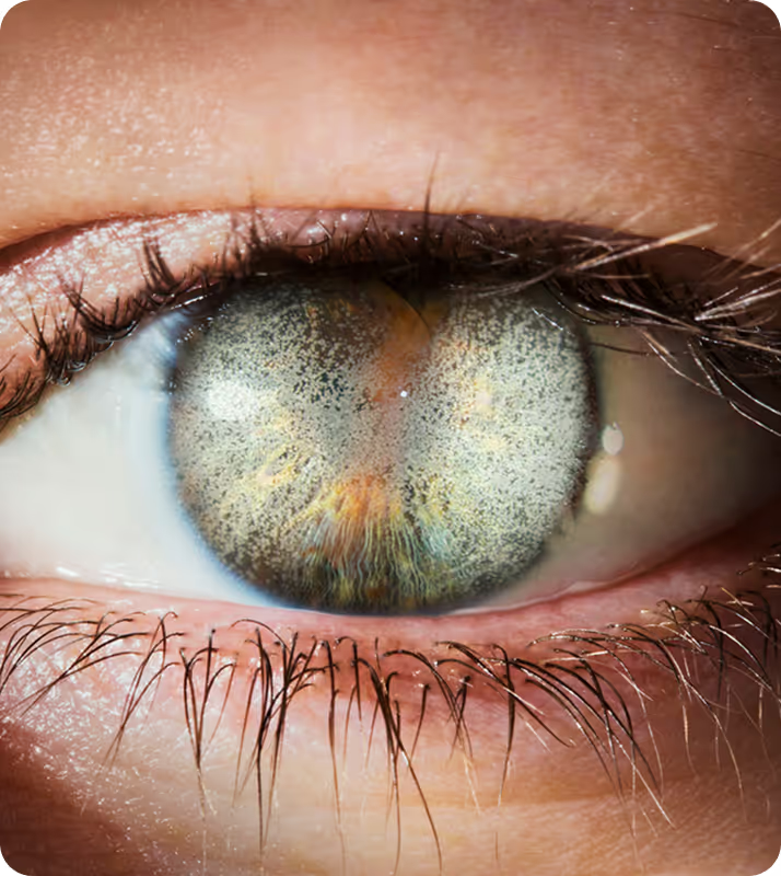
Corneal Degenerations. The cornea is the transparent outermost layer of the eye. Light must pass through the cornea to reach the pupil and retina. The cornea is crucial for refracting and focusing light—and therefore focusing your vision. As the eye’s outer layer, the cornea protects the eye from all kinds of infections and foreign objects and materials. The cornea is an extremely vital part of the eye and of your vision.
What is corneal degeneration?
Corneal degenerations are changes or gradual deteriorations in the tissue of the cornea. They can negatively impact the function of the cornea, limiting its ability to help the eye focus properly. Over time, corneal degenerations can cause loss of vision, eye pain, and other issues. The symptoms of corneal degeneration can vary significantly, as the condition has multiple varieties.
What causes corneal degeneration?
Corneal degenerations can be caused by disease or by aging. In most cases, degenerations have nothing to do with genetics. Age-related degenerations are known as “involutional” corneal degenerations, while degenerations caused by disease or exposure to certain conditions are “non-involutional.”
Involutional degenerations are significantly more common.
Multiple diseases and conditions can be contributing causes of corneal degenerations. For instance, band keratopathy degenerations are more common among patients with rheumatoid arthritis, glaucoma, syphilis, keratitis (corneal inflammation), and Crohn’s disease. Because these conditions have been linked with corneal degenerations, you should always provide a complete and accurate medical history to your eye doctor.
What are the symptoms of corneal degenerations?
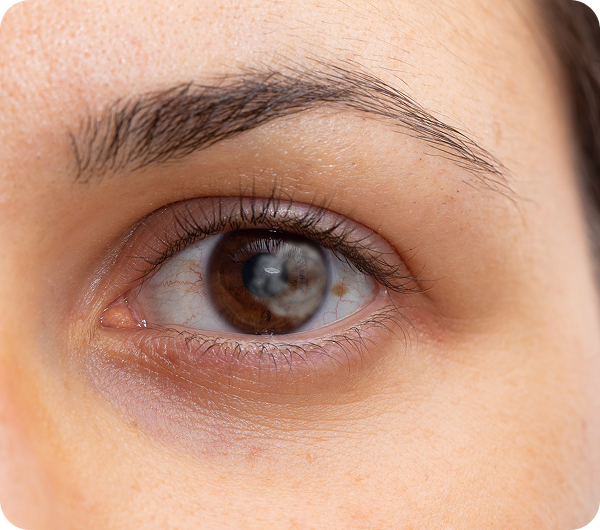
The symptoms of a corneal degeneration can vary depending on the type of degeneration. In most cases, you will experience both cosmetic and sight-related symptoms. Band keratopathy degenerations are characterized by calcium salt deposits building up in the cornea. This type of degeneration typically leads to severe vision loss, to the point where the patient has light perception but cannot detect much else. Salzmann’s nodular degeneration is characterized by blue nodules on the eye that cause extreme pain, and localized vision loss. And the White Limbal Girdle of Vogt—the most common type of degeneration caused by aging—is a thinning of the cornea’s periphery that causes visible changes along the corneal periphery but does not cause significant visual changes.
What is the difference between corneal degenerations and corneal dystrophies?
Corneal dystrophies are eye disorders that are passed down genetically. They can be progressive, leading many people to confuse them with corneal degenerations. Often, corneal dystrophies have earlier onset (around the age of 20) and eventually stabilize (around the age of 40). Dystrophies are caused by an accumulation of foreign material in the cornea. Usually, this buildup of material happens in just one of the corneal layers. However, most corneal dystrophies are bilateral, which means they occur in both eyes. Corneal dystrophies can result in eye pain or discomfort (including the feeling that there is a foreign body in the eye), as well as blurred vision and vision loss. As with corneal degenerations, the exact symptoms and severity of dystrophies depend on which type of dystrophy the patient is experiencing.
How are corneal degenerations treated?
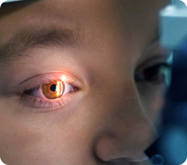
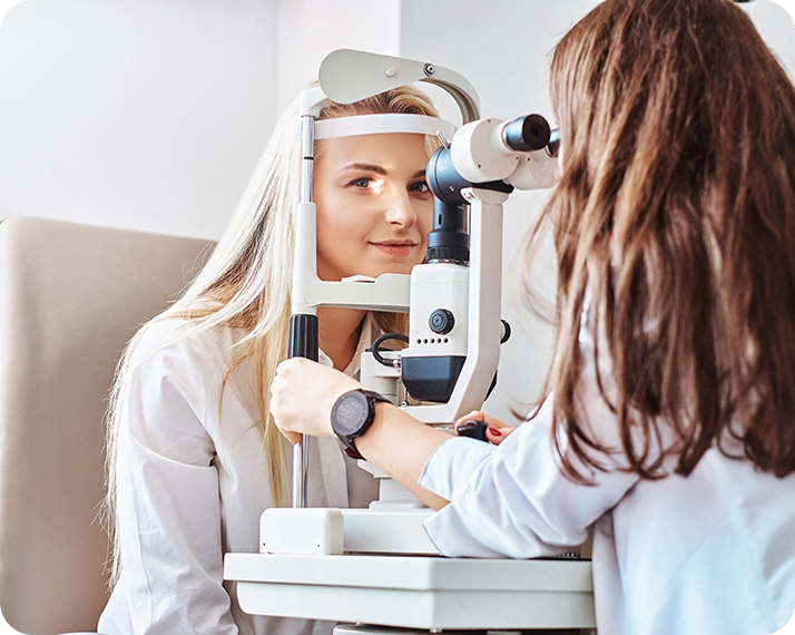
Treatments for corneal degenerations can vary depending on the exact diagnosis. To start, most eye doctors prescribe lubricating eye drops. Properly lubricating the eye can reduce the discomfort and pain associated with corneal degenerations. For conditions that have progressed further, an eye doctor might recommend surgery, such as a corneal transplant. If you suspect you have a corneal degeneration or dystrophy, make an appointment with your local eye doctor. An eye care practitioner will be able to give you a concrete diagnosis, explain the condition, and provide the best advice for ongoing care.
How Scleral Lenses Can Make Life Easier with Corneal Degeneration
Living with corneal degeneration means facing more than just blurry vision. Whether caused by keratoconus, pellucid marginal degeneration, or other thinning disorders, changes to the cornea can distort the way light enters the eye, making it difficult to drive, read, or recognize faces. Glasses often can't correct this distortion, and traditional contact lenses may feel uncomfortable or fail to provide stable vision. That’s where scleral lenses come in.
Unlike standard lenses, scleral lenses don’t rest on the sensitive cornea. Instead, they vault over it, sitting on the less sensitive white part of the eye (the sclera). This design creates a perfectly smooth optical surface, helping to neutralize the irregularities caused by corneal thinning or warping. For many patients, the result is a dramatic improvement in clarity; sharper, more stable vision that glasses simply can't provide.
In addition to vision correction, the fluid-filled space between the lens and the eye provides a protective cushion. This can shield the cornea from further irritation or mechanical trauma, especially in fragile or thinning areas. For people with advanced corneal conditions, this protective effect is not just a comfort feature, it’s essential!
What makes scleral lenses especially effective is how precisely customized they are. Using advanced imaging technology, each lens is individually designed to match the unique shape of your eye. This means they’re not only more comfortable, but also more stable, which translates to more stable vision throughout the day.
For many people with corneal degenerations, scleral lenses offer more than just a better way to see. They offer a safer, non-surgical option to improve daily function, preserve the integrity of the eye, and restore quality of life and confidence in everyday activities.